Ultrasound Scan Melbourne
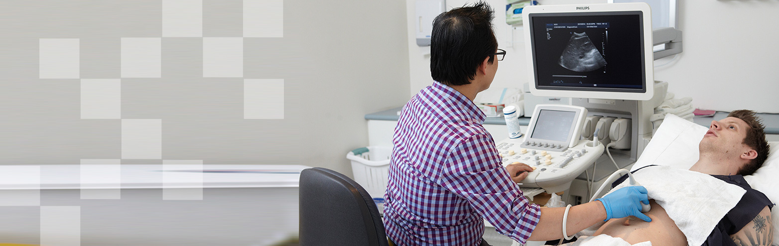
If you need to arrange an ultrasound scan, DiagnostiCare is a state-of-the-art clinic with experienced, supportive, and highly knowledgeable radiographers. From the initial consultation to post-scan analysis, our staff are here to provide a comfortable and respectful environment to all patients.
Utilising state-of-the-art technology, our team of highly trained technicians and radiologists provide accurate imaging for efficient diagnosis. This quality of service has made us a leading destination for ultrasound scans in Melbourne.
Get an Ultrasound at Our Keilor East Clinic
At DiagnostiCare, we can conduct a wide range of ultrasound scans in line with your personal requirements and needs. Our ultrasound scans cover a wide spectrum, from abdominal and pregnancy ultrasounds to testicular and thyroid analysis.
We understand that an ultrasound scan is more than just a procedure. It could potentially identify underlying health concerns that may need addressing. Therefore, we ensure that every scan we conduct is thorough, precise, and meticulous.
Using cutting-edge ultrasound technology, our trained professionals view your internal organs and systems, providing real-time imaging that aids in immediate and accurate diagnosis. Sometimes referred to as sonograms, ultrasounds utilise high-frequency sound waves that create live images from inside your body. Unlike X-ray imaging, there is no ionising radiation exposure associated with ultrasound scans.
Why Choose DiagnostiCare for an Ultrasound Scan in Melbourne
Here’s why DiagnostiCare is a trusted destination for your next ultrasound scan in Melbourne:
- Experienced Professionals: Our team comprises trained professionals, each bringing years of experience in conducting ultrasound scans accurately and efficiently.
- Patient-Centric Approach: At DiagnostiCare, every patient matters. We focus on providing superior comfort, dignity, and respect to every individual.
- State-of-Art Technology: We are equipped with cutting-edge ultrasound technology to provide high-resolution, real-time imaging for an accurate diagnosis.
Frequently Asked Questions About Ultrasound Scan Melbourne
DiagnostiCare, Melbourne excels in conducting a wide range of ultrasound scans as per your medical requirements. Our ultrasound scans cover a wide spectrum, including abdominal, pregnancy, testicular, and thyroid ultrasounds.
DiagnostiCare prioritises patient-centric care. From the initial consultation to post-scan follow-up, our professionals ensure a comfortable and respectful environment for our patients.
DiagnostiCare utilises state-of-the-art ultrasound technology for scanning. This technology provides high-resolution, real-time imaging, enabling significant accuracy in diagnostics.
DiagnostiCare, Melbourne boasts a team of highly experienced professionals, a patient-centric approach, and adherence to state-of-art technology. We are also a preferred ultrasound clinic in Keilor East, known for efficient and accurate services.
An ultrasound scan at DiagnostiCare, Melbourne gives you access to real-time imaging of your internal organs and systems. The procedure can identify underlying health concerns needing attention. Our ultrasound scans, also known as sonograms, utilise high-frequency sound waves to create live images without exposing you to ionising radiation associated with X-ray imaging.