MRI Melbourne
At DiagnostiCare, we provide high-quality Magnetic Resonance Imaging (MRI) services in Melbourne. With cutting-edge technology and a team of dedicated professionals, our team provides accurate diagnoses and personalised patient care.
Our top-of-the-line MRI machines utilise a potent magnetic field and radio waves to create detailed images of the body’s internal structure, aiding in the identification of various health issues.
We accept referrals and in possession of a full MRI liscence, which allows us to bulk bill designated Specialist and GP referred studies.. Please contact us now to book an MRI appointment.
Comprehensive MRI Services in Melbourne
Our fully trained and experienced radiologists specialise in providing MRI scans which are highly sensitive and can produce precise images of soft-tissues, bones, and nerves in your body. Whether you need a brain, spine, or joint MRI, we have the expertise and the technology required to provide you with accurate results.
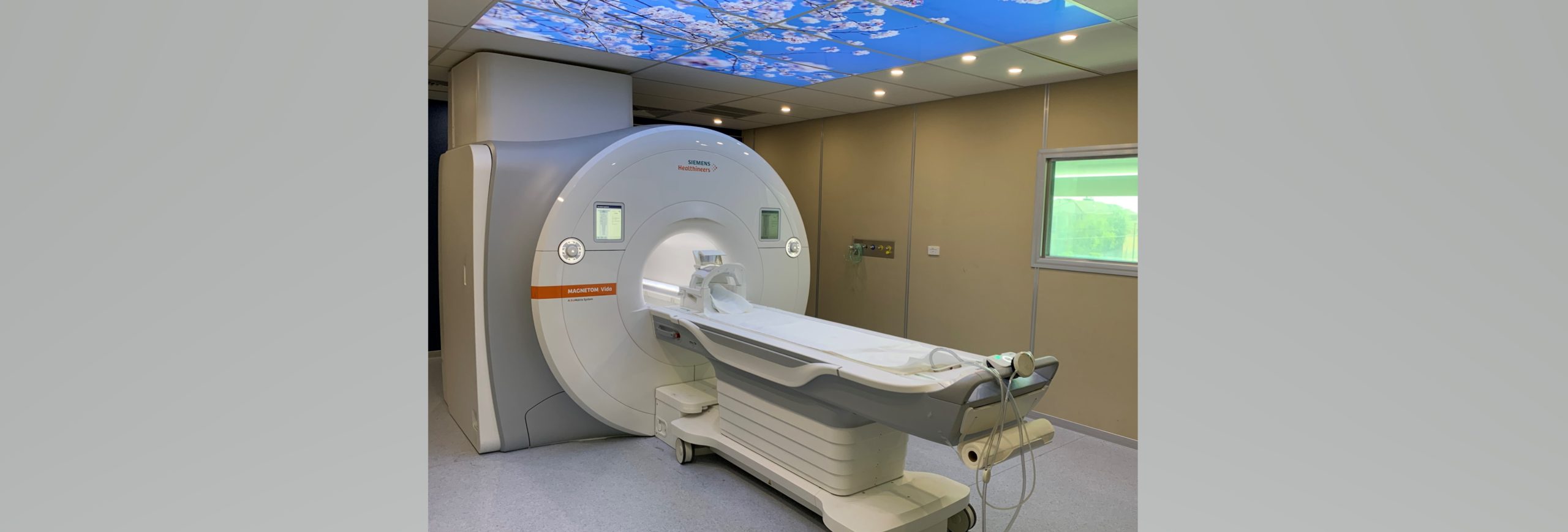
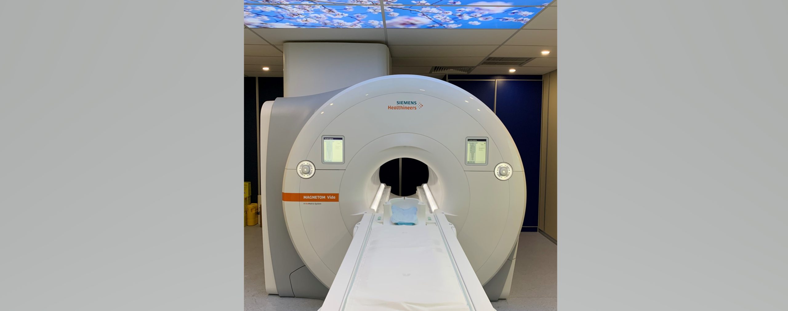
Our Melbourne-based MRI service follows a stringent procedure. Patients will be given a robe to wear and positioned on an MRI compatible table. The table is then slid into our wide bore MRI scanner, a doughnut-shaped device deep.
Prostate MRI Melbourne
Regular prostate scans are incredibly important for all men of a certain age. At DiagnostiCare, we conduct accurate prostate MRI scans at our Melbourne clinic. This process is handled by highly knowledgeable, friendly, and professional radiographers.
Prostate MRI is a non-invasive procedure that provides highly accurate images, aiding doctors in the early detection and treatment of prostate cancer. Our entire process is handled with the utmost care, keeping our patients’ comfort and ease as our priority.
MRI Services in Keilor East
Serving the local community of Keilor East, our comprehensive MRI services are available to all who need it.We run 2 MRI scanners:a 3 Tesla and a 1.5 Tesla magnet, both of which are fully specced and have the latest Siemens Boost AI technology installed. With our commitment to patient care and our state-of-the-art technology, we offer an MRI experience that is second to none. Our trained staff will guide you through the process, making sure you are comfortable and aware of the procedure.
The Benefits of an MRI Scan
MRI scans provide images for a variety of conditions, including, but not limited to, tumours, bleeding, infection, blood vessel diseases, or injury. It is renowned for its potential to detect conditions early, which can significantly impact the overall health outcomes for patients. A clear advantage of MRI scans is that they involve no radiation exposure, offering safer diagnostic imaging, especially for individuals requiring frequent scanning.
Why Choose DiagnostiCare for MRI Scans in Melbourne?
Choosing DiagnostiCare for your MRI needs means opting for a high-quality, accurate, and compassionate service. Here are a few reasons why DiagnostiCare stands out:
- Our team of radiologists are subspeciality trained and have years of experience in the field.
- We use the latest, and tope of the range equipment and technology to ensure high-quality imaging.
- We prioritise patient comfort and aim to create a stress-free experience.
- Our services are more likely to be bulk billed, as we have a full MRI licence, and if not bulk billable, then our rates are very competitive when compared to outher radiology practicess.
- We are conveniently located and cater to Melbourne and surrounding suburbs, with lots of available parking..
A sincere commitment to our patients’ health and well-being powers our work. We guarantee timely, accurate, and reliable diagnostic imaging services to meet your healthcare needs. contact us today on (03) 9337 8288 to schedule your appointment.
Frequently Asked Questions About MRI Scans in Melbourne
DiagnostiCare in Melbourne offers a wide range of MRI scans. We specialise in brain, Paediatric,spine, joint,cardiacprostate MRI scans, amongst others. Our state-of-the-art equipment can create precise images of various parts of the body, aiding in accurate diagnosis and treatment.
At DiagnostiCare, patient comfort is one of our top priorities. The MRI scanner used is doughnut-shaped and only 1.2 metres deep, ensuring most scans are performed with the patient’s head well out of the scanner to alleviate any claustrophobia concerns. One of our scanners is fitted with the latest Siemens In-View in-bore entertainment system, which allows you to watch any movie or video material of your choice, while you are having the scan.Members of our staff are also at hand to guide patients through the process and ensure they are comfortable.
Our prostate MRI service in Melbourne plays a significant role in prostate health. It is a non-invasive procedure that provides highly accurate images. This aids doctors in the early detection and treatment of prostate conditions, including prostate cancer. The comprehensive procedure is performed with the utmost care, aiming to ensure patient comfort.
Yes, we extend our excellent MRI services to the local community of Keilor East. Our dedicated team is committed to providing quality care, ensuring that our patients are comfortable and well-informed throughout the process.
DiagnostiCare should be your preferred choice for MRI in Melbourne due to a number of reasons. We have a full MRI licence, and this allows us to bulk bill the majority of referrals from specialists, and for certain specified indications from general practitioners.Our radiologists are well trained and experienced, and we use the latest technology to ensure high-quality imaging. Additionally, we prioritise patient comfort and offer our services at affordable rates. DiagnostiCare is conveniently located and caters to residents in Melbourne and its surrounding suburbs.